Figures 28–33
Download original image
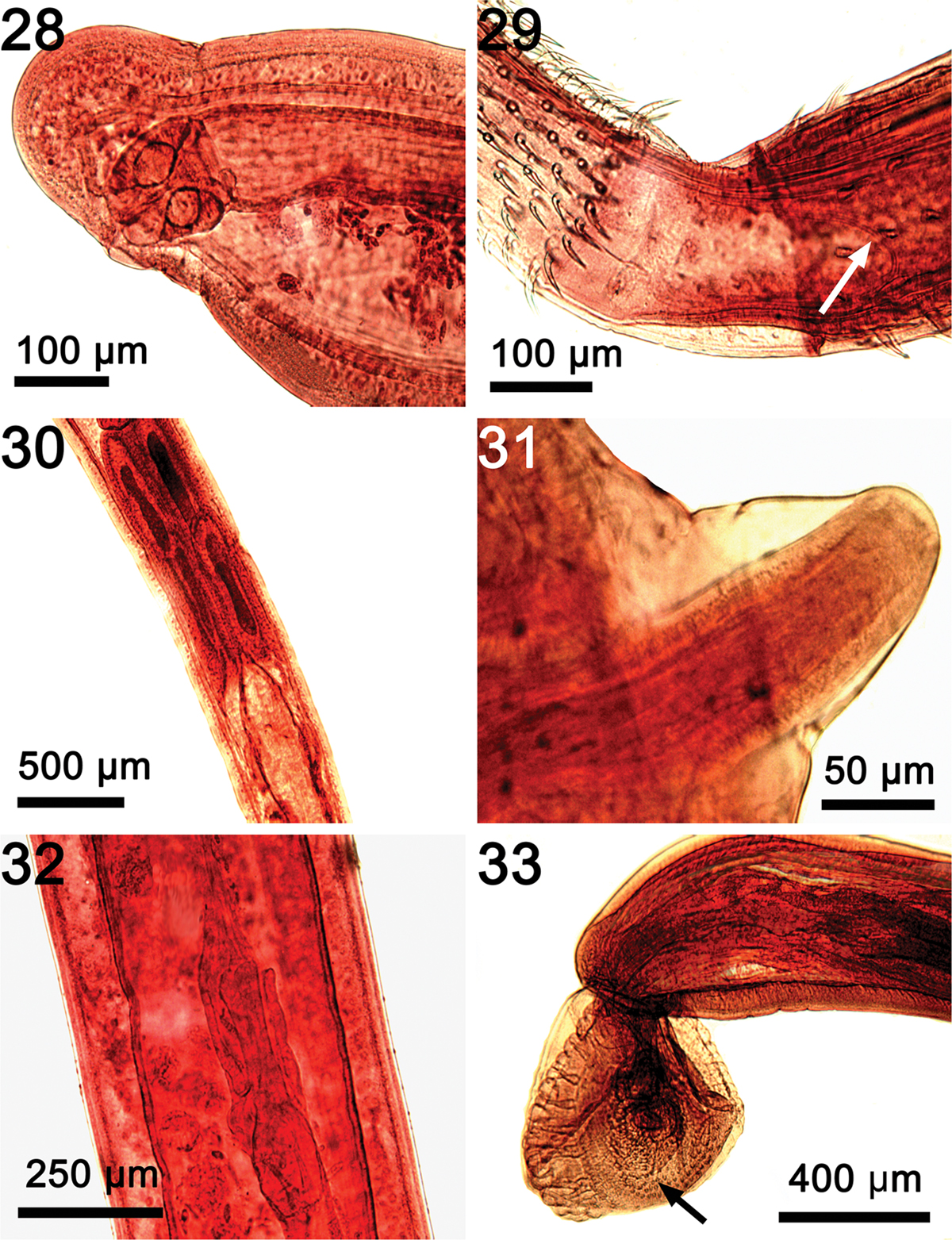
Microscopical images of some internal structures as seen in their natural state not readily demonstrable in line drawings of specimens of Rhadinorhynchus laterospinosus from Auxis rochei and Auxis thazard in the Pacific Ocean off Vietnam. (28) A sub-ventral vagina at the constriction of posterior end of trunk of a female. (29) The posterior loop of a thin sac (arrow) emerging from the insertion of the proboscis receptacle at the base of the proboscis. (30) The four tubular cement glands with their long nuclei just anterior to Saefftigen’s pouch. (31) The penis emerging from the bursa of one specimen. (32) Uterine bell in one female. Note the unequal sides of the bell. (33) The posterior end of one male showing the bursa with rings of sensory papillae (arrow).
Current usage metrics show cumulative count of Article Views (full-text article views including HTML views, PDF and ePub downloads, according to the available data) and Abstracts Views on Vision4Press platform.
Data correspond to usage on the plateform after 2015. The current usage metrics is available 48-96 hours after online publication and is updated daily on week days.
Initial download of the metrics may take a while.