Figures 22–27
Download original image
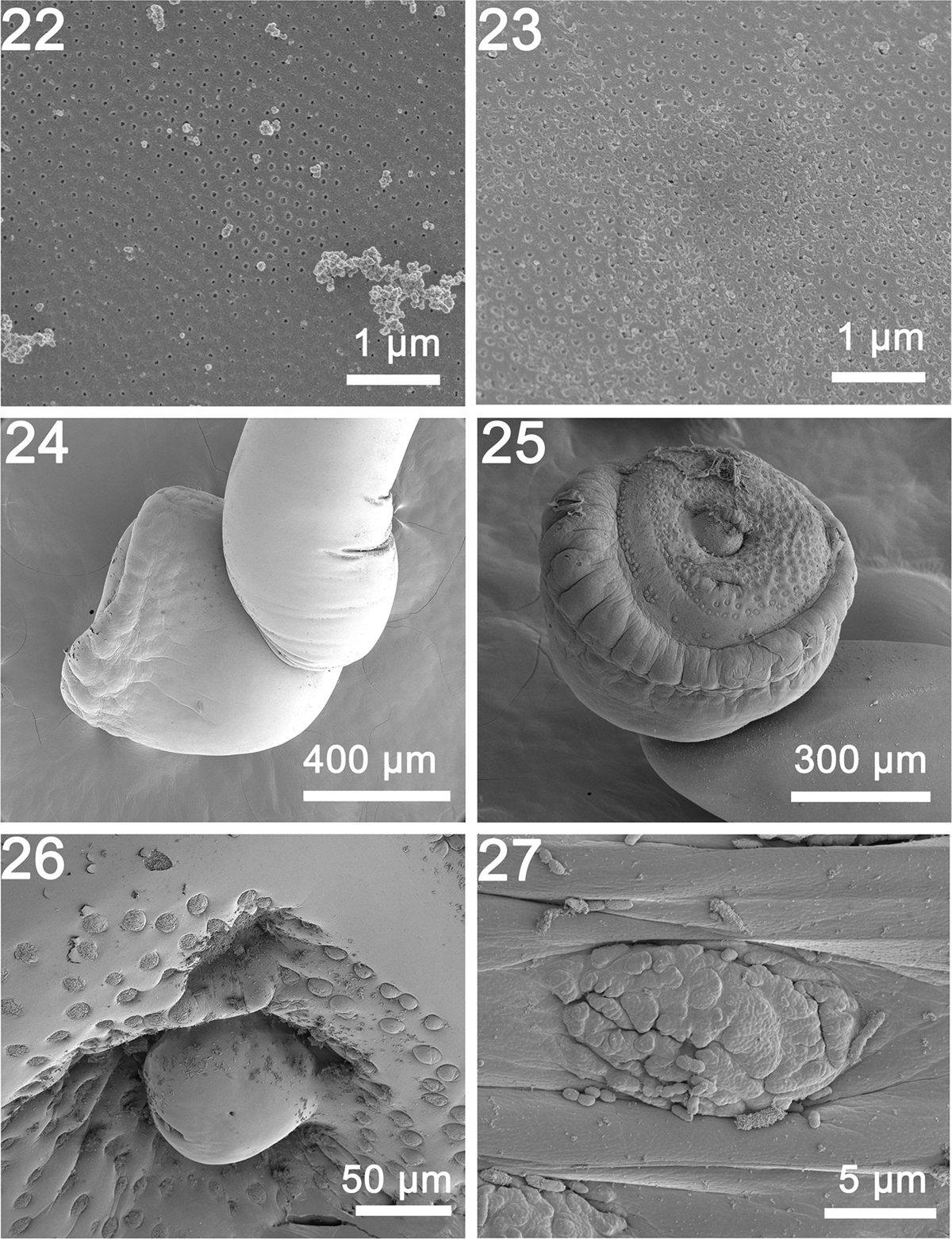
SEM of specimens of Rhadinorhynchus laterospinosus from Auxis rochei and Auxis thazard in the Pacific Ocean off Vietnam. (22, 23) Microspores from the middle and posterior parts of the trunk, respectively. Note the different density and diameter of the pores, also compared with Figure 18 related to differential absorption rates. (24) A lateral view of the bursa. (25) A ventrolateral view of a bursa showing its thick muscular margin and the organization of the outer circle and the central cluster of sensory papillae. (26) A high magnification of the center of the bursa showing the terminal genitalia surrounded by close circles of sensory papillae. This organization is species-specific. (27) A higher magnification of one sensory papilla made up of small units embedded in elliptic depression.
Current usage metrics show cumulative count of Article Views (full-text article views including HTML views, PDF and ePub downloads, according to the available data) and Abstracts Views on Vision4Press platform.
Data correspond to usage on the plateform after 2015. The current usage metrics is available 48-96 hours after online publication and is updated daily on week days.
Initial download of the metrics may take a while.