Figures 1–9
Download original image
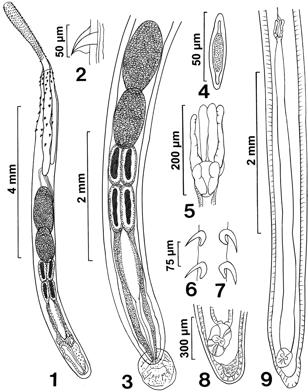
Line drawings of whole mounted specimens of Rhadinorhynchus laterospinosus from Auxis rochei and Auxis thazard in the Pacific Ocean off Vietnam. (1) A paratype male showing the anteriorly enlarged proboscis, the distribution of trunk spines within the range of the long receptacle, and the posterior distribution of the reproductive system. (2) A posterior ventral trunk spine of a female specimen. (3) Detailed male reproductive system. Note the large tubular giant nuclei of the cement glands and the posterior extension of the cement gland ducts surrounding Saefftigen’s pouch anteriorly. (4) A ripe egg. (5) Detail of the uterine bell of the female specimen shown in Figure 9. Note the inner paired rod-like structures. (6, 7) Dorsal (Fig. 6) and ventral (Fig. 7) hooks at the mid proboscis of a female specimen. Note differences in the thickness, length, and curvature of dorsal vs. ventral hooks. (8) Detail of the vagina from Figure 9. Note the inner muscular plug lining of the posterior tip of the trunk. (9) A complete female reproductive system characterized by the long and wide uterus.
Current usage metrics show cumulative count of Article Views (full-text article views including HTML views, PDF and ePub downloads, according to the available data) and Abstracts Views on Vision4Press platform.
Data correspond to usage on the plateform after 2015. The current usage metrics is available 48-96 hours after online publication and is updated daily on week days.
Initial download of the metrics may take a while.