Figure 1
Download original image
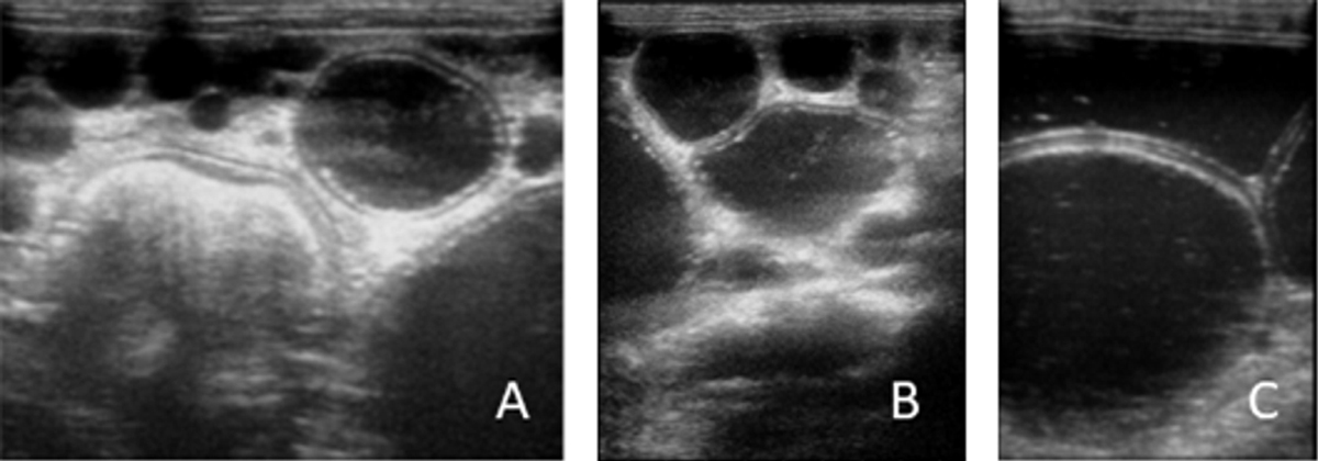
Abdominal ultrasound images. Hydatids either attached to the omentum (A) or apparently free in the peritoneal cavity (B) with anechoic content and delimited by a hyperechoic rim. Particular of a large peritoneal hydatid with evident bilaminated structure of the wall appearing as a double echogenic line separated by a hypoechogenic space (C).
Current usage metrics show cumulative count of Article Views (full-text article views including HTML views, PDF and ePub downloads, according to the available data) and Abstracts Views on Vision4Press platform.
Data correspond to usage on the plateform after 2015. The current usage metrics is available 48-96 hours after online publication and is updated daily on week days.
Initial download of the metrics may take a while.