Figure 9
Download original image
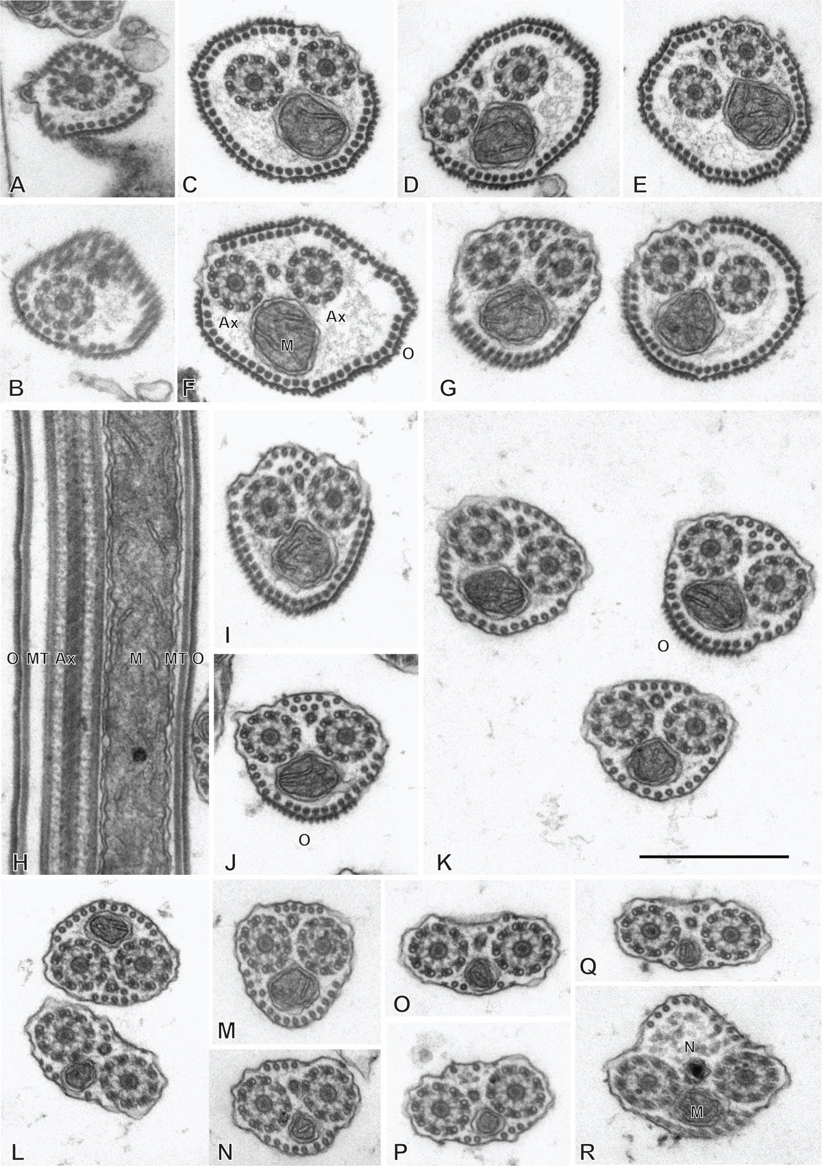
Spermatozoon of Rajonchocotyle emarginata, anterior part. A-G and I-R, transverse sections, in antero-posterior sequence; H, longitudinal section. A, B, anteriormost part of spermatozoon: one axoneme, one centriolar derivative, peripheral row of microtubules and ornamentation on membrane. C-G, region of spermatozoon with membrane ornamentation. Microtubules generally as a single row of peripheral parallel units, two axonemes and section of mitochondrion. G shows together a typical section (right) and an intermediary section (left) with ornamentation only on ventral side of spermatozoon. H, longitudinal section in region with membrane ornamentation, showing axoneme with typical trepaxonematan 9 + ”1” structure, mitochondrion with very regular diameter, peripheral microtubules and external ornamentation on cell membrane (microtubules and ornamentation visible on both sides). I-K, various sections at the limit of region with ornamentation and region without ornamentation and additional microtubules. A section of mitochondrion is visible in all sections; a section of nucleus appears, with very small diameter. L-R, sections in the region posterior to membrane ornamentation, showing decreasing number of external microtubules and progressive appearance of section of nucleus. Scale in K, valid for all figures: 500 nm.
Current usage metrics show cumulative count of Article Views (full-text article views including HTML views, PDF and ePub downloads, according to the available data) and Abstracts Views on Vision4Press platform.
Data correspond to usage on the plateform after 2015. The current usage metrics is available 48-96 hours after online publication and is updated daily on week days.
Initial download of the metrics may take a while.