Figures 20-25
Download original image
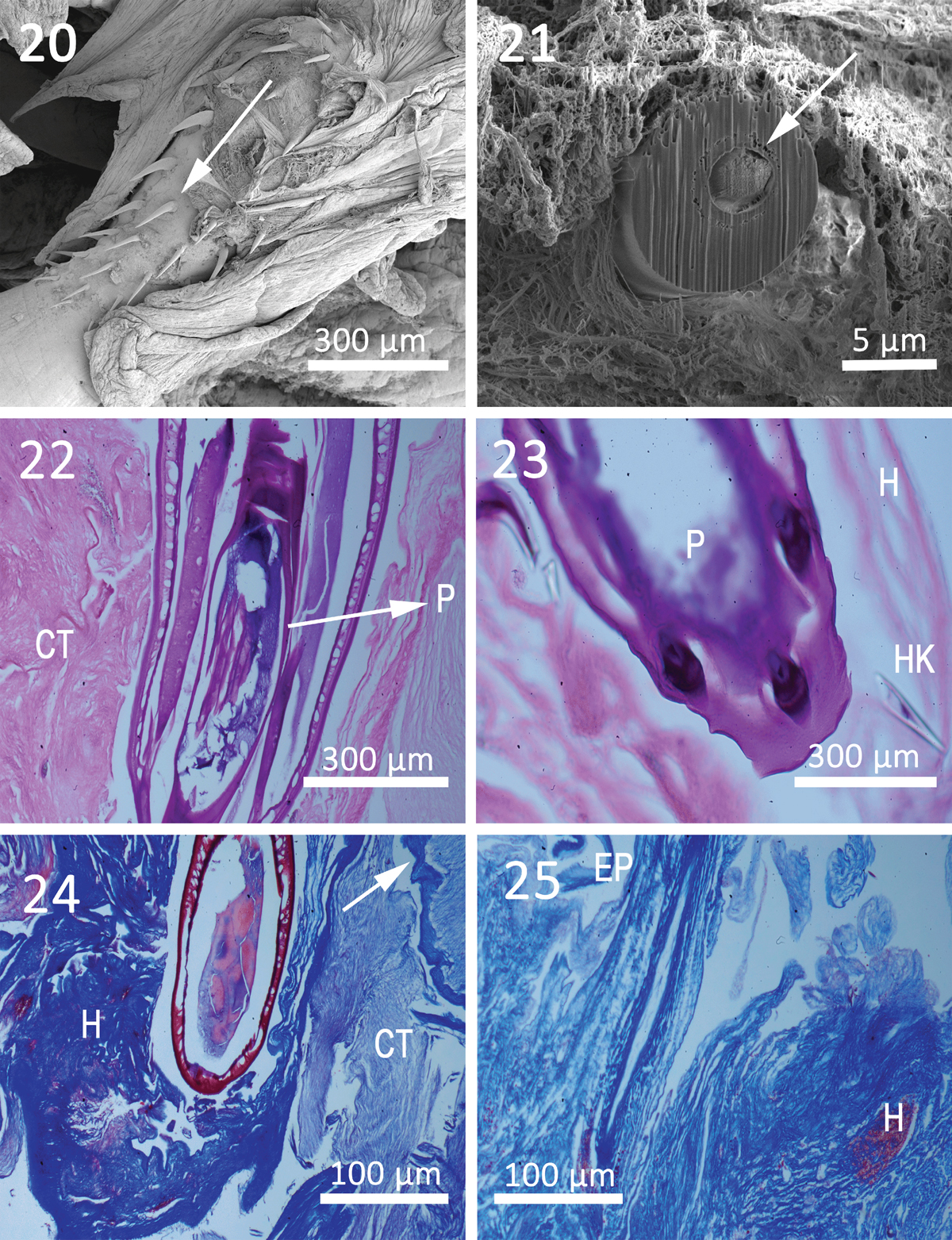
Histopathology of Cavisoma magnum in the intestinal track of Mugil cephalus from the Arabian Gulf. 20. SEM of attached worm; note hooks (arrow) on the proboscis of a worm. This image shows the gross pathology and the extreme damage to host intestinal tissue. 21. Gallium-cut hook (arrow) from proboscis of attached worm. Note host connective tissue surrounding worm. 22. Proboscis (P) of a worm. Host connective tissue (CT) is visible with remnants of the intestinal epithelium next to the worm. 23. Proboscis (P) of a worm with sections of worm hooks (HK) and host tissue (H) surrounding worm. 24. Trichrome preparation of infected host tissue (H) section and worm body are visible. Note remnants of host intestine (arrow). 25. Area where worm had infected the host tissue (H). Note ports of hemorrhaged blood caused by worm penetration and remnants of the host intestinal epithelium (EP).
Current usage metrics show cumulative count of Article Views (full-text article views including HTML views, PDF and ePub downloads, according to the available data) and Abstracts Views on Vision4Press platform.
Data correspond to usage on the plateform after 2015. The current usage metrics is available 48-96 hours after online publication and is updated daily on week days.
Initial download of the metrics may take a while.