Figure 3.
Download original image
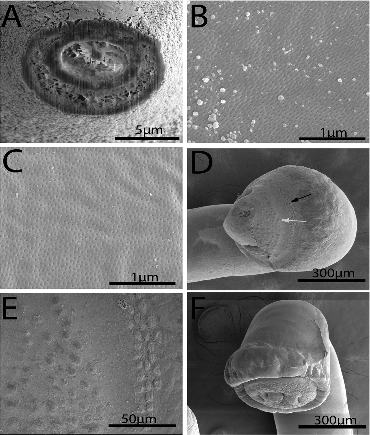
SEM image of specimens of Rhadinorhynchus oligospinosus from Scomber japonicus and Trachurus murphyi from off the Pacific coast of Peru. (A) A gallium cut section of a trunk spine at its base showing the differentiation between its cortical and core layers and associated spaces. (B) Micropores at the anterior part of the trunk. (C) Micropores at the Bmid-section of the trunk. Note the difference in the diameter and distribution of micropores at different trunk regions. (D) A dorsal view of the bursa showing the distribution of the outer zone (black arrow) and the inner zone (white arrow) of sensory receptors. (E) A larger magnification of sensory receptors in the outer and inner zones. (F) A ventro-dorsal perspective of a bursa showing its thick muscular margin and its terminal gonopore and appendage.
Current usage metrics show cumulative count of Article Views (full-text article views including HTML views, PDF and ePub downloads, according to the available data) and Abstracts Views on Vision4Press platform.
Data correspond to usage on the plateform after 2015. The current usage metrics is available 48-96 hours after online publication and is updated daily on week days.
Initial download of the metrics may take a while.