Figure 4.
Download original image
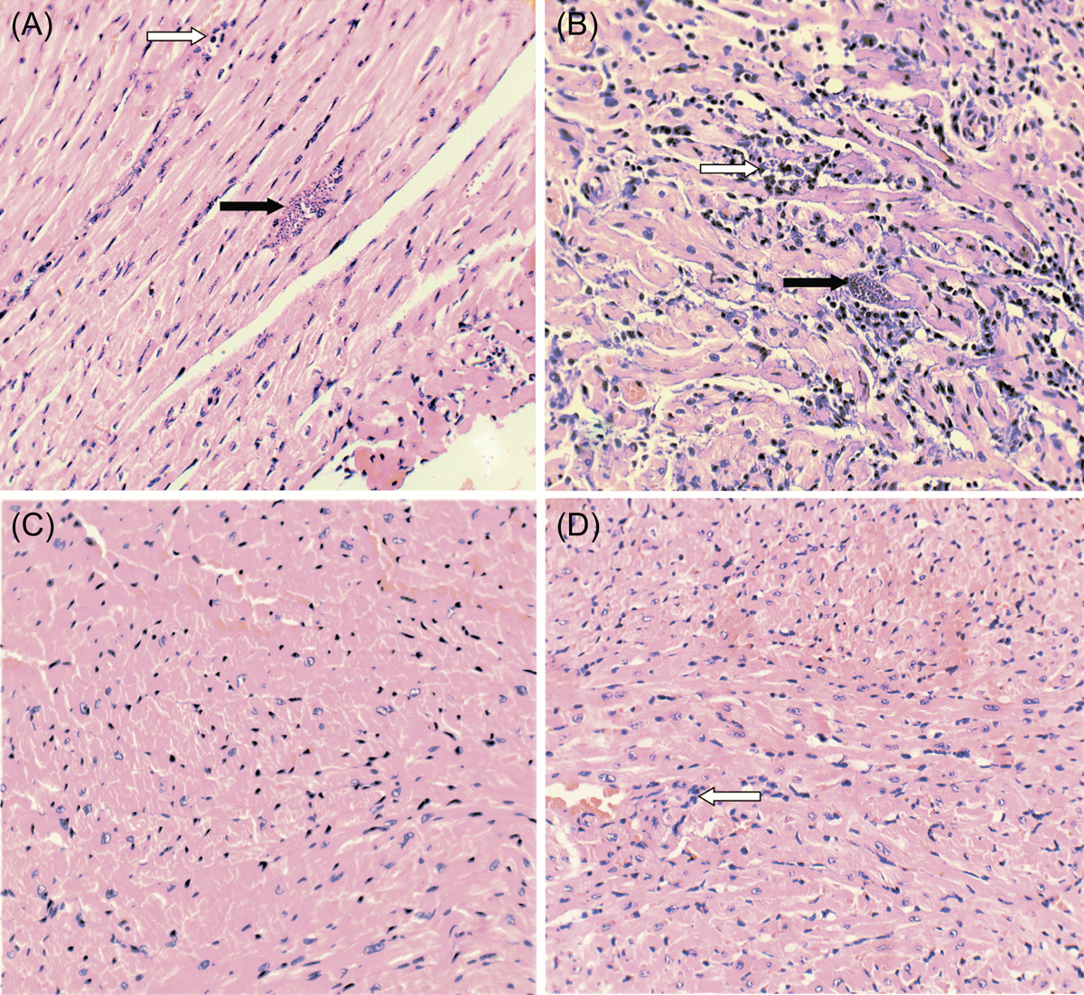
Histological analysis of heart tissue sections from acutely Trypanosoma cruzi infected mice, treated with astaxanthin and/or nifurtimox. Heart tissue sections from the left ventricle were processed on the day animals died (either due to infection or when euthanized on day 60 post-infection). Tissue sections (5 μm) were stained with hematoxylin-eosin. Representative micrographs are shown for mice from the following groups: (A) G1 (Tc); (B) G2 (Tc/ASTX); (C) G5 (saline solution); (D) G8 (ASTX). The micrograph from G5 could represent all groups from G3 to G7; all of them were considered histologically normal. Black arrow, amastigote nests; white arrow, lymphoplasmacytic infiltrate. (400× amplification).
Current usage metrics show cumulative count of Article Views (full-text article views including HTML views, PDF and ePub downloads, according to the available data) and Abstracts Views on Vision4Press platform.
Data correspond to usage on the plateform after 2015. The current usage metrics is available 48-96 hours after online publication and is updated daily on week days.
Initial download of the metrics may take a while.