Figure 4.
Download original image
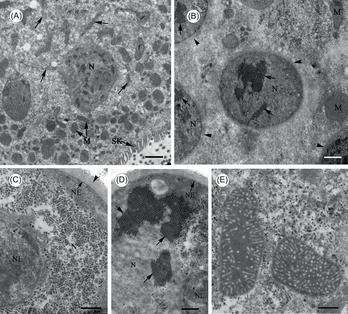
Transmission electron microscope images of Cepedea longa, to show fine structures of the nuclei and mitochondria within the endoplasm. (A) Cross section observed at low magnification, to show numerous thin bundles of microfilaments (arrow) dispersed in the endoplasm between nuclei (N) and mitochondria (M). SK = somatic kinetosomes. Scale bar = 20 μm. (B)–(D) Cross section of the nuclei (N), to show the nuclear envelope (NE) covered by a thick layer of microfibrils (arrowhead) and some unknown microtubular structures (arrow) in the nucleoplasm. NL = nucleolus. Scale bar in B = 10 μm, in C and D = 5 μm. (E) Thin section shows mitochondria having tubular cristae at periphery with an amorphic appearance. Scale bar = 5 μm.
Current usage metrics show cumulative count of Article Views (full-text article views including HTML views, PDF and ePub downloads, according to the available data) and Abstracts Views on Vision4Press platform.
Data correspond to usage on the plateform after 2015. The current usage metrics is available 48-96 hours after online publication and is updated daily on week days.
Initial download of the metrics may take a while.