Figure 3.
Download original image
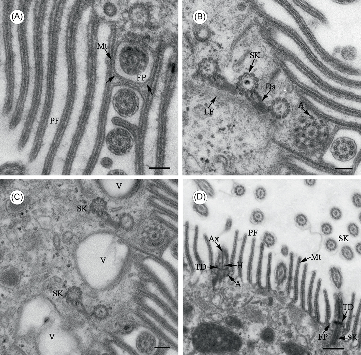
Transmission electron microscope images of Cepedea longa, to show fine structures of the somatic flagella. (A) Tangential section of a somatic kinety, to show fibrillar elements (arrow) between cortical microtubules (Mt) and around the membrane of each flagellar pit (FP). PF = pellicular folds. Scale bar = 2.5 μm. (B)–(C) Cross section through several kineties, to show somatic kinetosomes (SK) linked by desmoses (Ds) and sometimes interposed by vacuoles (V) just beneath the cell surface. A = kinetosomal arms. Scale bar = 2.5 μm. (D) Longitudinal section of kinetosomes, to show detailed fine structures. The axosome (Ax) is embedded in the proximal margin of transitional discs (TD), with curving arms (A) extending out and up. H = transitional helix, Mt = microtubules, SK = somatic kineties, PF = pellicular folds, FP = flagellar pit. Scale bar = 5 μm.
Current usage metrics show cumulative count of Article Views (full-text article views including HTML views, PDF and ePub downloads, according to the available data) and Abstracts Views on Vision4Press platform.
Data correspond to usage on the plateform after 2015. The current usage metrics is available 48-96 hours after online publication and is updated daily on week days.
Initial download of the metrics may take a while.