Figure 2.
Download original image
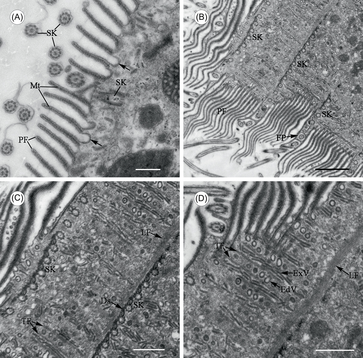
Transmission electron microscope images of Cepedea longa, to show fine structures of the somatic cortex. (A) Section tangent to cell surface, to show pellicular folds (PF) supported by ribbons of microtubules (Mt). Some coated vesicles are fused with the plasma membrane and seen as invaginations (arrow). SK = somatic kinetosomes. Scale bar = 5 μm. (B) Section passing parallel to cell surface, to show pellicular folds (PF) interposing between somatic kineties (SK). FP = flagellar pit. Scale bar = 20 μm. (C)–(D) Selected enlargement of Figure 2A, to show a developed fibrillar skeletal system in the somatic cortex. Longitudinal microfibrils (LF) border the somatic kineties (SK) joined to each other by desmoses (Ds) on the left side, with transversal fibrils (TF) running perpendicular to kinetal long axes and framing the ribs of the cortical vesicles: globular endocytotic vesicles (EdV) and elongated exocytotic vesicles (ExV). Scale bar = 10 μm.
Current usage metrics show cumulative count of Article Views (full-text article views including HTML views, PDF and ePub downloads, according to the available data) and Abstracts Views on Vision4Press platform.
Data correspond to usage on the plateform after 2015. The current usage metrics is available 48-96 hours after online publication and is updated daily on week days.
Initial download of the metrics may take a while.