Figure 1.
Download original image
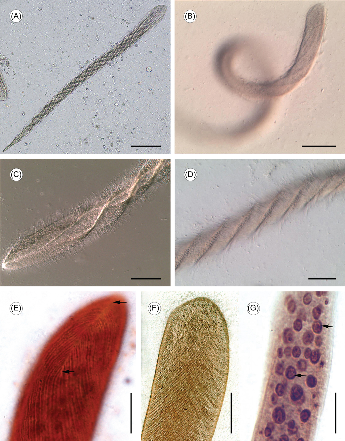
Light microscope images of Cepedea longa. (A) Overview of the living specimens, to show general form, greatly elongated and cylindrical, with the anterior extremity broader and the posterior end pointed. Scale bar = 100 μm. (B) Living specimens, to show C. longa thickly flagellated and often coils when swimming. Scale bar = 100 μm. (C)–(D) Living specimens, to show body surface twisting and giving a spiral appearance. Scale bar = 50 μm. (E) Specimens stained with ammoniacal silver, to show the falx (arrow) and somatic kineties branching off from each side. Scale bar = 25 μm. (F) Specimens stained with silver nitrate, to show somatic kineties follow a sigmoid course from anterior to posterior end of the cell. Scale bar = 25 μm. (G) Specimens stained with ammoniacal silver, to show the organism possessing a large amount of spherical or ellipsoidal nuclei (arrow). Scale bar = 25 μm.
Current usage metrics show cumulative count of Article Views (full-text article views including HTML views, PDF and ePub downloads, according to the available data) and Abstracts Views on Vision4Press platform.
Data correspond to usage on the plateform after 2015. The current usage metrics is available 48-96 hours after online publication and is updated daily on week days.
Initial download of the metrics may take a while.