Figures 5–8.
Download original image
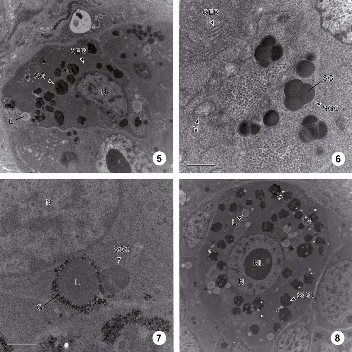
Stages 3 and 4 of vitellogenesis in C. metoecus. (5) General observation of a vitelline cell at the third stage of maturation. Scale bar = 1 μm. (6) The third stage of vitellocyte maturation showing the coalescence of single shell globules into a cluster surrounded by a membrane. Scale bar = 1 μm. (7) Cytoplasm of a vitelline cell at the third stage of maturation containing shell globule cluster and saturated lipid droplets, surrounded by glycogen granules. Stained according to the Thiéry method. Scale bar = 1 μm. (8) Electron micrograph of a vitelline cell at the fourth stage of maturation filled with shell globule clusters and lipid droplets. Scale bar = 1 μm. G: glycogen particle; GER: granular endoplasmic reticulum; L: lipid droplet; M: mitochondrion; N: nucleus; Nl: nucleolus; SG: shell globule; SGC: shell globule cluster.
Current usage metrics show cumulative count of Article Views (full-text article views including HTML views, PDF and ePub downloads, according to the available data) and Abstracts Views on Vision4Press platform.
Data correspond to usage on the plateform after 2015. The current usage metrics is available 48-96 hours after online publication and is updated daily on week days.
Initial download of the metrics may take a while.