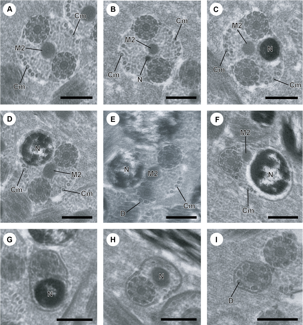
Figure 2.
Download original image
A–I. Mature spermatozoon of Atractotrema sigani in regions IV–V. (A) Cross-section in proximal part of region IV exhibiting the second mitochondrion (M2). Cm, cortical microtubules. Note the decrease in the maximum number of cortical microtubules (about 9) compared to region III. (B–D) Consecutive cross-sections showing the simultaneous presence of the second mitochondrion and the nucleus (N) which increases progressively in size. (E) Disorganization of the first axoneme resulting into doublets of microtubules. D, doublet of microtubules. (F) Distal part of region IV with only the second axoneme, the second mitochondrion and few cortical microtubules (about 4). (G, H) Cross-section showing only the second axoneme, the nucleus and its progressive reduction in size. (I) Cross-section of the posterior spermatozoon tip, where the nucleus disappears and only one axoneme is still observed. Scale in μm: (A–I), 0.3.
Current usage metrics show cumulative count of Article Views (full-text article views including HTML views, PDF and ePub downloads, according to the available data) and Abstracts Views on Vision4Press platform.
Data correspond to usage on the plateform after 2015. The current usage metrics is available 48-96 hours after online publication and is updated daily on week days.
Initial download of the metrics may take a while.