Figures 1–12.

Download original image
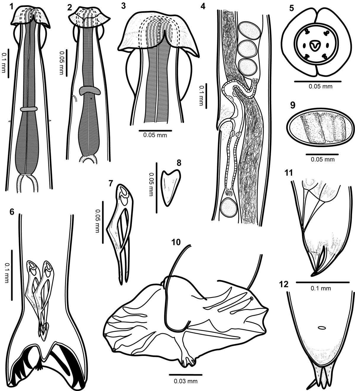
Torrestrongylus tetradorsalis n. sp. 1: Anterior end of male, ventral view, showing the esophagus and the relative positions of the nerve ring and deirids; note separation of cuticular projection in anterior vesicle. 2: Anterior end of female, lateral view showing the relative position of the nerve ring, deirid, and excretory pore; note continuous cuticular expansion in anterior vesicle. 3: Detail of the cephalic vesicle showing the anterior half with the lateral projections forming an umbrella and the posterior half with “handles of a pitcher” appearance. 4: Ovejector of female showing flap in anterior lip of vulva, vagina vera, infundibulum, sphincters, and uterine branches containing mature eggs. 5: Face view of male, showing arrangement of papillae and cuticular expansions of the anterior cephalic vesicle. 6: Posterior end of male, showing the relative position of spicules and gubernaculum. 7: Spicule. 8: Gubernaculum. 9: Egg. 10: Caudal bursa showing bursal arrangement and bifurcation of dorsal ray. 11: Lateral view of tail of female. 12: Ventral view of tail of female.
Current usage metrics show cumulative count of Article Views (full-text article views including HTML views, PDF and ePub downloads, according to the available data) and Abstracts Views on Vision4Press platform.
Data correspond to usage on the plateform after 2015. The current usage metrics is available 48-96 hours after online publication and is updated daily on week days.
Initial download of the metrics may take a while.


