Figures 120–128.
Download original image
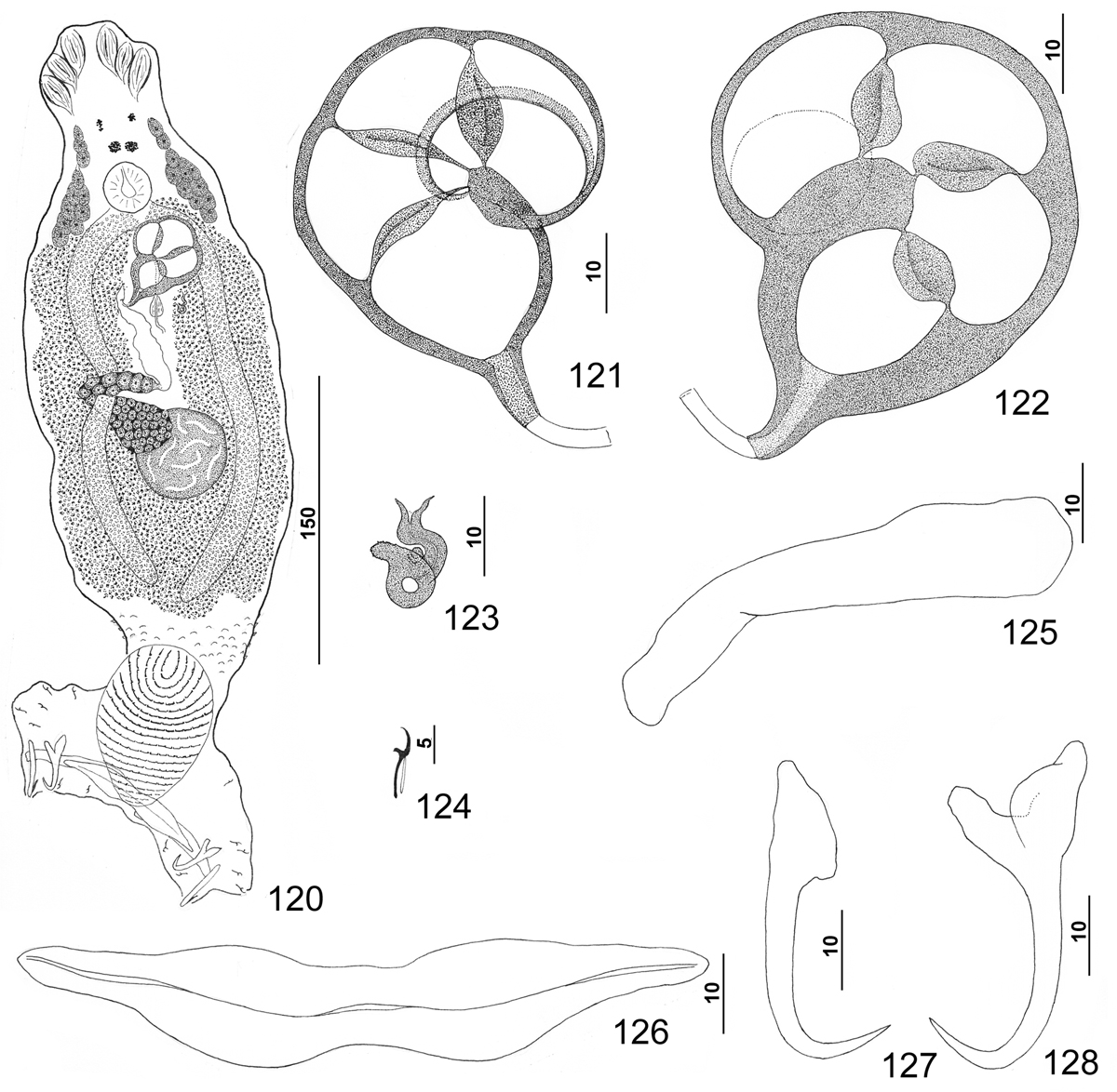
Pseudorhabdosynochus williamsi n. sp. from rock hind Epinephelus adscensionis. 120: whole mount (composite, ventral view; dorsal squamodisc and dorsal anteromedial haptoral lobe not shown); 121: male copulatory organ (dorsal view) with thin chamber walls; 122: male copulatory organ (ventral view) with thick walls of the chambers; 123: vaginal sclerite (ventral view); 124: hook; 125: right dorsal bar (ventral view); 126: ventral bar; 127: dorsal anchor; 128: ventral anchor.
Current usage metrics show cumulative count of Article Views (full-text article views including HTML views, PDF and ePub downloads, according to the available data) and Abstracts Views on Vision4Press platform.
Data correspond to usage on the plateform after 2015. The current usage metrics is available 48-96 hours after online publication and is updated daily on week days.
Initial download of the metrics may take a while.