Figure 3.
Download original image
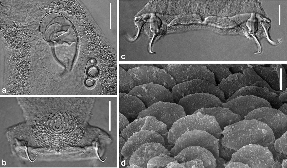
Pseudorhabdosynochus jeanloui n. sp. from Paranthias colonus. a–c, brightfield differential interference contrast microscope images. a, detail of quadriloculate organ and sclerotized vagina, holotype, Hoyer, ventral view. b, haptor, detail of ventral squamodisc and ventral hamuli, holotype, Hoyer, ventral view. c, haptor, detail of ventral and dorsal hamuli and bars, paratype, Hoyer, ventral view. d, scanning electron microscope image, detail of rodlet rows of a squamodisc, ventral view. Scale bars: a–b, 40 μm; c, 20 μm; d, 2 μm.
Current usage metrics show cumulative count of Article Views (full-text article views including HTML views, PDF and ePub downloads, according to the available data) and Abstracts Views on Vision4Press platform.
Data correspond to usage on the plateform after 2015. The current usage metrics is available 48-96 hours after online publication and is updated daily on week days.
Initial download of the metrics may take a while.