Figure 1.
Download original image
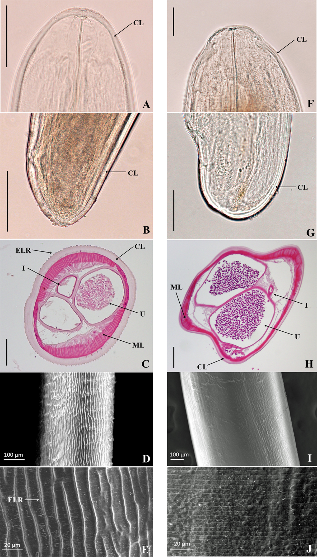
Morphological images of female adults of the Dirofilaria species isolated from the present case (left column, A–E) and Dirofilaria immitis (right column, F–J). A, F: direct images of the cephalic parts under an optical microscope. B, G: direct images of the caudal parts under an optical microscope. C, H: cross-sectional tissue sections (hematoxylin and eosin stain). D, I: low-magnification images of the body surfaces under a scanning electron microscope (SEM). E, J: high-magnification images of the body surfaces under SEM. Scales bars: A, B, C, F, G, 100 μm; H, 200 μm. CL, Cuticular Layer; ELR, External Longitudinal Ridge; I, Intestine; ML, Muscular Layer; U, Uterus.
Current usage metrics show cumulative count of Article Views (full-text article views including HTML views, PDF and ePub downloads, according to the available data) and Abstracts Views on Vision4Press platform.
Data correspond to usage on the plateform after 2015. The current usage metrics is available 48-96 hours after online publication and is updated daily on week days.
Initial download of the metrics may take a while.