Figure 1.
Download original image
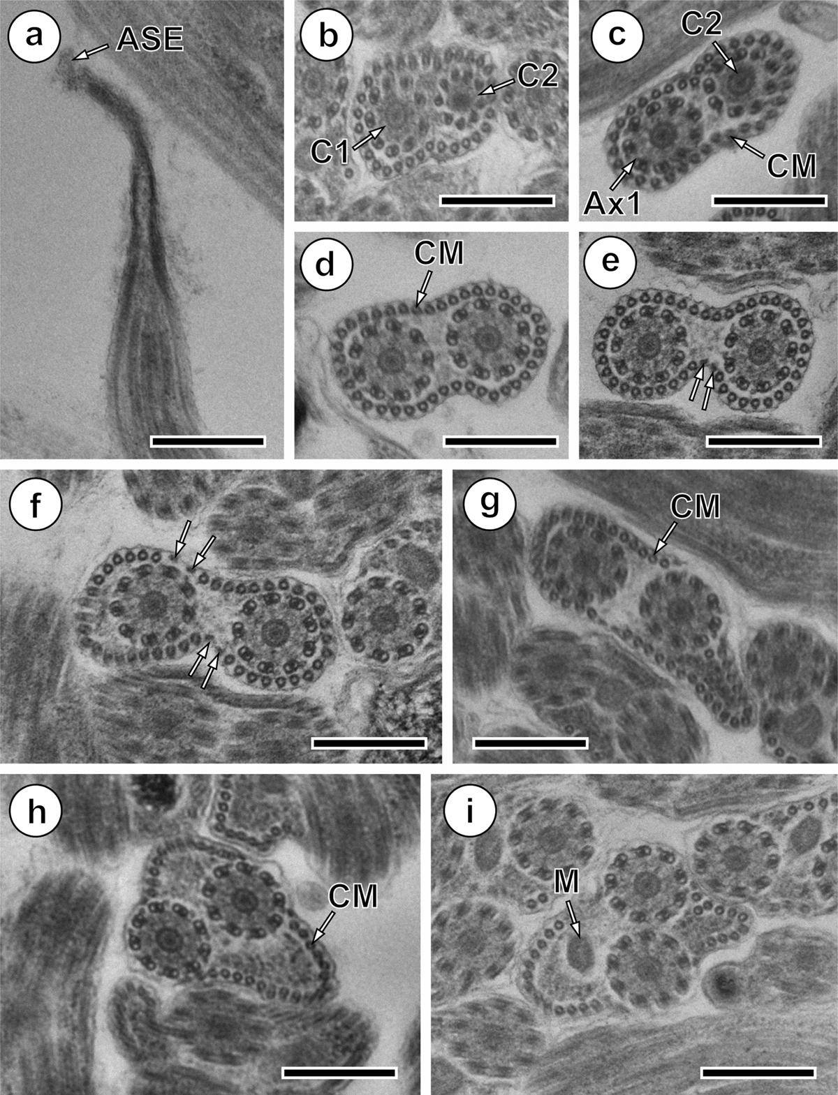
Mature spermatozoon of Collyricloides massanae. (a) Sharp morphology of the anterior spermatozoon extremity (ASE). (b, c) Cross-sections in Region I showing two centrioles (C1 and C2) corresponding to both axonemes and continuous layer of submembranous cortical microtubules (CM). Ax1, first axoneme. (d) Cross-section in which both axonemes are already formed and surrounded by a continuous layer of parallel cortical microtubules. (e, f) Consecutive cross-sections in the middle part of Region I exhibiting two and four attachment zones (arrows), interrupting the continuous layer of cortical microtubules. (g–i) Posterior part of Region I showing in cross-sections both axonemes and cortical microtubules organised into two fields separated by the four attachment zones. The appearance of the mitochondrion is also noticeable (M). Scale in μm: (a), 0.5; (b–i), 0.3.
Current usage metrics show cumulative count of Article Views (full-text article views including HTML views, PDF and ePub downloads, according to the available data) and Abstracts Views on Vision4Press platform.
Data correspond to usage on the plateform after 2015. The current usage metrics is available 48-96 hours after online publication and is updated daily on week days.
Initial download of the metrics may take a while.