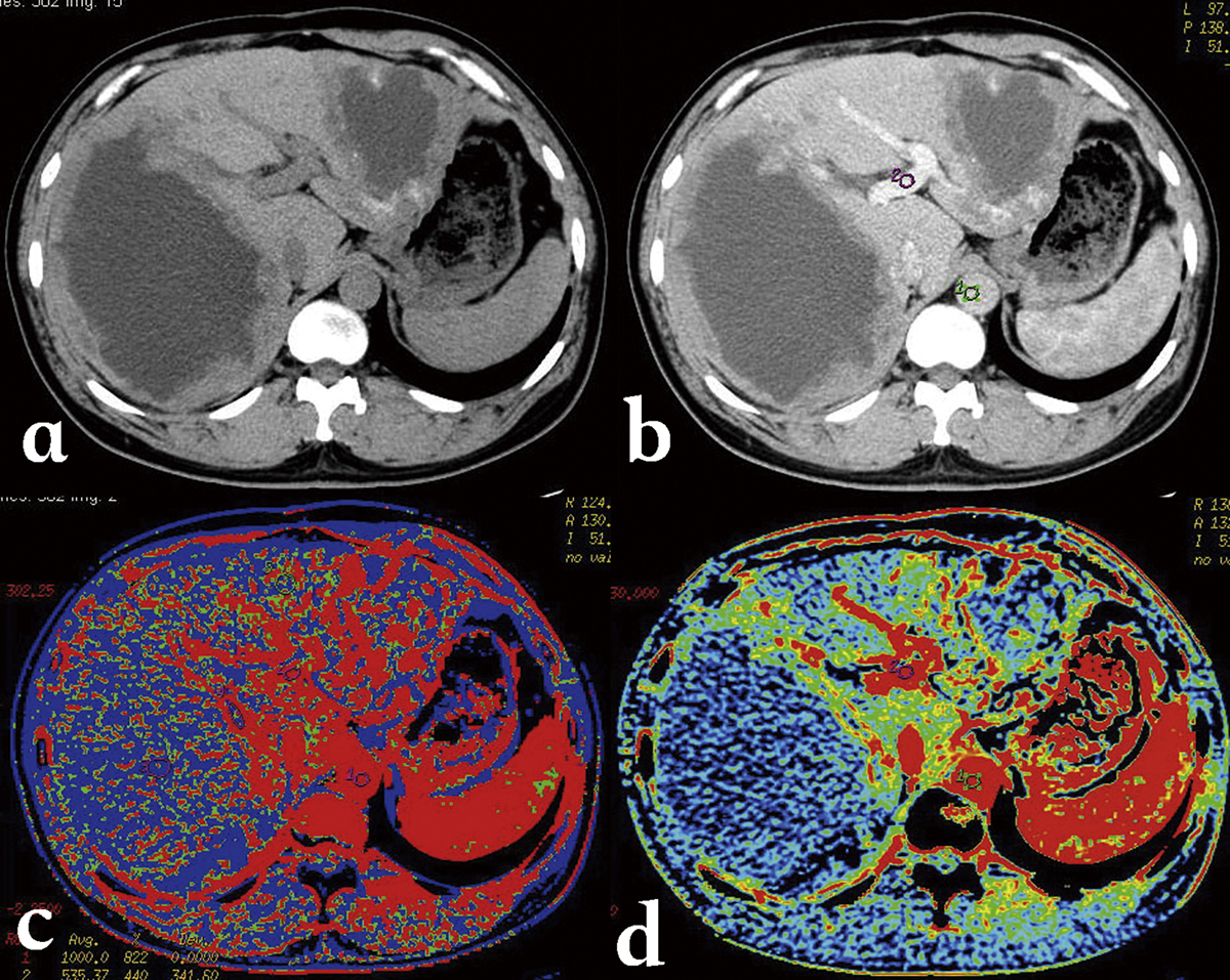
Figure 8.
Download original image
Alveolar echinococcosis in a 34-year-old man. (a) CT plain scan shows a cyst-solid mixed hepatic mass involving both lobes of liver with irregular margin and necrosis areas of low density in the center. (b) Post-contrast phase shows a non-enhancing, hypo-attenuating lesion. (c) Blood flow map, and (d) blood volume map, at CT perfusion, show the micro-circulation in the marginal area of AE.
Current usage metrics show cumulative count of Article Views (full-text article views including HTML views, PDF and ePub downloads, according to the available data) and Abstracts Views on Vision4Press platform.
Data correspond to usage on the plateform after 2015. The current usage metrics is available 48-96 hours after online publication and is updated daily on week days.
Initial download of the metrics may take a while.