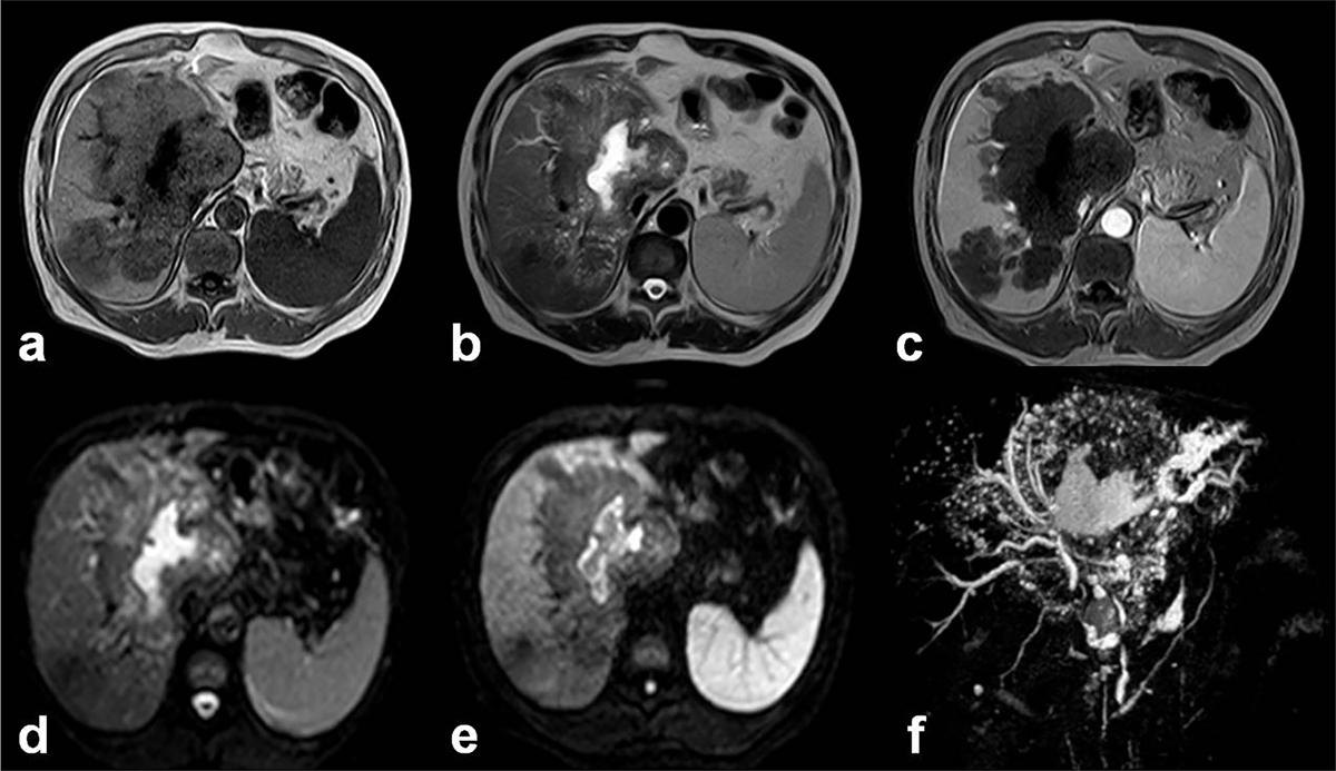
Figure 4.
Download original image
Alveolar echinococcosis in a 55-year-old man. (a) Axial unenhanced T1-weighted Magnetic Resonance (MR) image shows infiltrating diffused mass with hypo-intense signal in the right and left lobes of the liver. (b) Axial unenhanced T2-weighted MR image shows heterogeneous signal. The high signal intensity corresponds to small cystic and necrotic components of mass lesion. (c) Axial enhanced T1-weighted image with gadolinium shows no enhancement in the mass lesion. (d, e) Diffusion-weighted MR images obtained with b value of 0 s/mm2 and 600 s/mm2. Hypo-intense signal characterizes the cystic and necrotic components of the lesion whereas hyper-intense signal seen in the central necrotic part reveals restricted diffusion due to bacterial superinfection. (f) MR Cholangiopancreatography of the liver shows several small cystic and necrotic components of the mass lesion with dilated right hepatic duct, constricted upper part of common bile duct, and mildly dilated pancreatic duct.
Current usage metrics show cumulative count of Article Views (full-text article views including HTML views, PDF and ePub downloads, according to the available data) and Abstracts Views on Vision4Press platform.
Data correspond to usage on the plateform after 2015. The current usage metrics is available 48-96 hours after online publication and is updated daily on week days.
Initial download of the metrics may take a while.