Figure 2.
Download original image
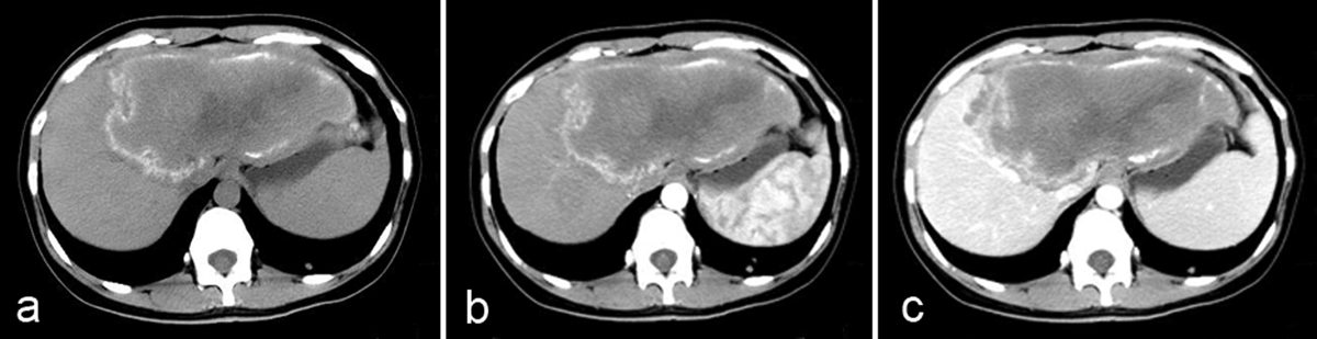
Alveolar echinococcosis in a 28-year-old woman. (a) Axial unenhanced CT image shows an infiltrative tumor-like hepatic mass involving both lobes of the liver with irregular margin and heterogeneous contents including hyper-attenuating foci of calcification scattered peripherally and an area of hypo-attenuation corresponding to necrosis and parasitic tissue centrally. (b) Post-contrast arterial phase shows a non-enhancing, hypo-attenuating lesion. (c) Post-contrast portal-venous phase shows a faint enhancement of fibro-inflammatory components surrounding the parasitic pseudo-cyst.
Current usage metrics show cumulative count of Article Views (full-text article views including HTML views, PDF and ePub downloads, according to the available data) and Abstracts Views on Vision4Press platform.
Data correspond to usage on the plateform after 2015. The current usage metrics is available 48-96 hours after online publication and is updated daily on week days.
Initial download of the metrics may take a while.