Figure 11.
Download original image
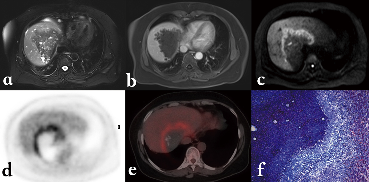
Alveolar echinococcosis in a 45-year-old patient with hepatic alveolar echinococcosis (HAE). (a) T2-weighted MR images showed a heterogeneous mass: note the multiple vesicles in the lesion, which showed a well-defined border on enhanced T1WI due to the absence of enhancement of the lesion itself (b). (c) Diffusion-weighted MR images revealed a half circular hyper-intensity area at the lesion’s border with the normal liver parenchyma, which was confirmed to be metabolically active by PET (d) and PET/CT (e). (f) HAE peripheral area was composed of severe fibrosis combined with a large number of inflammatory cells on the histopathological sections (Masson staining; ×100).
Current usage metrics show cumulative count of Article Views (full-text article views including HTML views, PDF and ePub downloads, according to the available data) and Abstracts Views on Vision4Press platform.
Data correspond to usage on the plateform after 2015. The current usage metrics is available 48-96 hours after online publication and is updated daily on week days.
Initial download of the metrics may take a while.