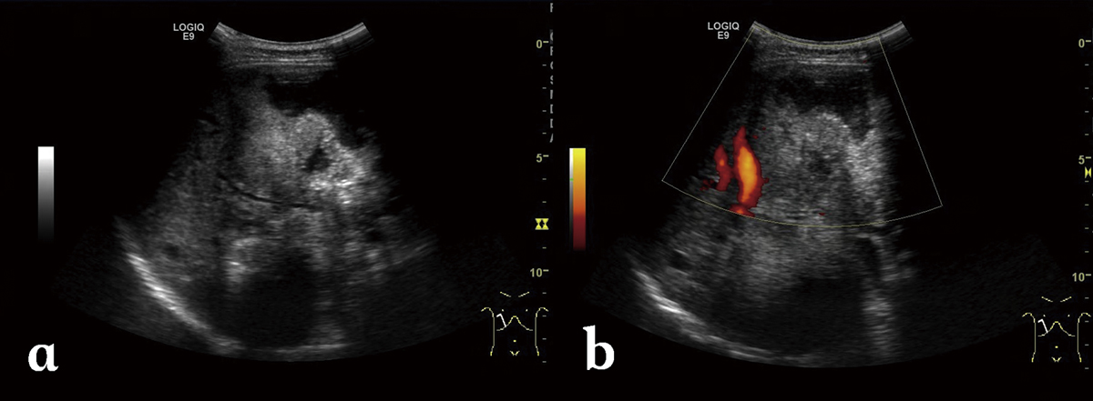
Figure 1.
Download original image
Alveolar echinococcosis in a 30-year-old woman. (a) Abdominal gray-scale US image shows an irregular type heterogeneous mass lesion with no clear boundary in the right lobe of the liver, containing anechoic pseudo-cystic lesion and hyperechoic foci of calcification. (b) Color Doppler US image shows no obvious blood flow signal while cystic duct is constricted.
Current usage metrics show cumulative count of Article Views (full-text article views including HTML views, PDF and ePub downloads, according to the available data) and Abstracts Views on Vision4Press platform.
Data correspond to usage on the plateform after 2015. The current usage metrics is available 48-96 hours after online publication and is updated daily on week days.
Initial download of the metrics may take a while.