Figure 2.
Download original image
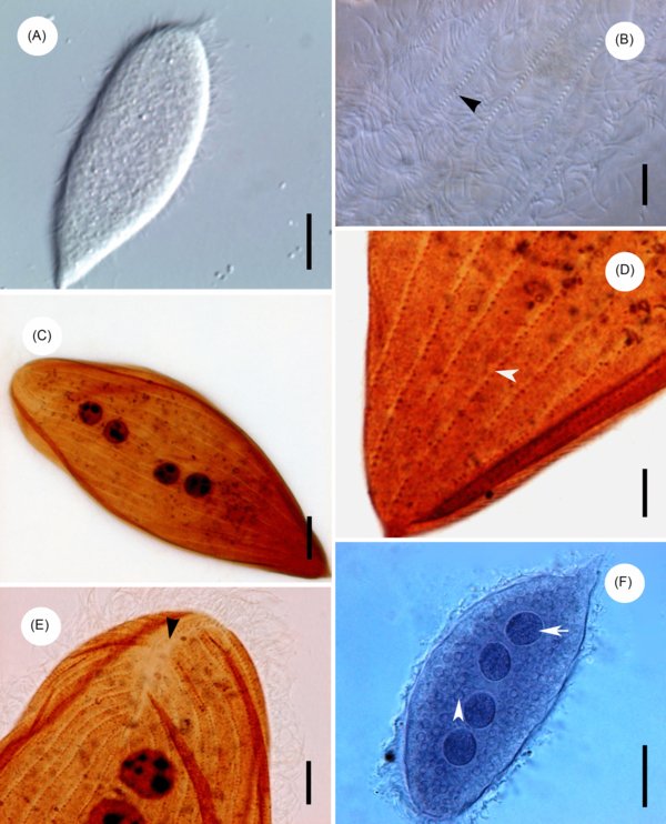
Light microscope images of Protoopalina pingi Nie, 1935. (A) Living specimens, showing the normal trophozoites of P. pingi. Scale bar = 20 μm. (B) Living specimens, showing the flagella covering the body (arrowhead). Scale bar = 5 μm. (C) Specimens stained with Protargol, showing the somatic kineties and the nuclei with distributed nucleoli. Scale bar = 10 μm. (D) Specimens stained with Protargol, showing the somatic kineties in the posterior extremity (arrowhead). Scale bar = 5 μm. (E) Specimens stained with Protargol, showing the falx region in the anterior extremity (arrowhead). Scale bar = 5 μm. (F) Specimens stained with Heidenhain’s haematoxylin, showing the nuclei (arrow) and the corpuscles of uneven size (arrowhead). Scale bar = 20 μm.
Current usage metrics show cumulative count of Article Views (full-text article views including HTML views, PDF and ePub downloads, according to the available data) and Abstracts Views on Vision4Press platform.
Data correspond to usage on the plateform after 2015. The current usage metrics is available 48-96 hours after online publication and is updated daily on week days.
Initial download of the metrics may take a while.