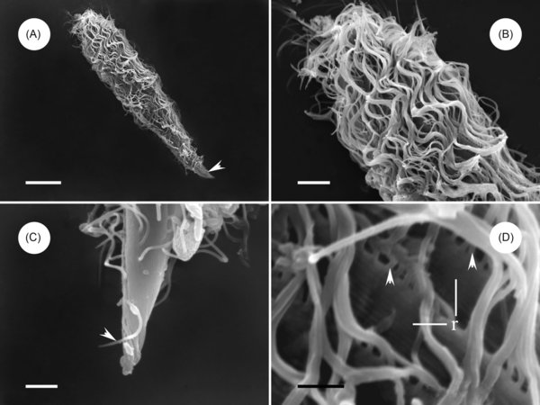
Figure 1.
Download original image
Scanning electron microscope images of Protoopalina pingi Nie, 1935. (A) Overview of P. pingi, showing many fused flagella over the body. Scale bar = 20 μm. (B) Anterior end of P. pingi, showing the densely flagellated body surface. Scale bar = 5 μm. (C) Caudal tip of P. pingi, showing the flagella (arrowhead) in the region barren of flagella. Scale bar = 2.5 μm. (D) The flagella are arranged in the ridge, showing the proximal ends of the flagella (arrowhead) and ridge (r). Scale bar = 1.5 μm.
Current usage metrics show cumulative count of Article Views (full-text article views including HTML views, PDF and ePub downloads, according to the available data) and Abstracts Views on Vision4Press platform.
Data correspond to usage on the plateform after 2015. The current usage metrics is available 48-96 hours after online publication and is updated daily on week days.
Initial download of the metrics may take a while.