Figure 1.

Download original image
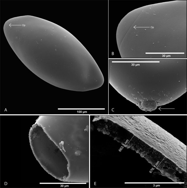
Scanning electron micrographs of Protopolystoma xenopodis egg features. (A) Fully embryonated egg, operculum visible (← Op). (B) Operculum becoming visible as the egg develops (← Op). (C) A residual structure on the non-opercular side of the egg. (D) An empty egg shell after the oncomiracidium has left. (E) Egg shell indicating the thickness of an individual parasite egg shell at the opercular opening.
Current usage metrics show cumulative count of Article Views (full-text article views including HTML views, PDF and ePub downloads, according to the available data) and Abstracts Views on Vision4Press platform.
Data correspond to usage on the plateform after 2015. The current usage metrics is available 48-96 hours after online publication and is updated daily on week days.
Initial download of the metrics may take a while.


