Figure 1.
Download original image
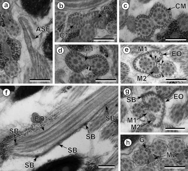
Spermatozoon of Pleurogenes claviger, anterior and middle parts (Regions I and II). (a) Longitudinal section of the anterior spermatozoon extremity (ASE). (b, c) Consecutive sections of the anterior area of the sperm cell showing the two centrioles (C1 and C2) and the presence of the complete submembranous layer of parallel cortical microtubules (CM). (d) Cross-section showing the appearance of the first mitochondrion (M1) and two attachment zones (arrowheads). (e–g) Longitudinal and cross-sections of the ornamented area of the spermatozoon. Note the presence of external ornamentation of the plasma membrane (EO), spine-like bodies (SB) and the second mitochondrion (M2). In this area, the cortical microtubules are organized into two fields separated by four attachment zones (arrowheads). M1, first mitochondrion. (h) Cross-section of the middle part of the spermatozoon (Region II) showing the appearance of granules of glycogen (G) and the presence of only the second mitochondrion (M2). Scales in μm: 0.3.
Current usage metrics show cumulative count of Article Views (full-text article views including HTML views, PDF and ePub downloads, according to the available data) and Abstracts Views on Vision4Press platform.
Data correspond to usage on the plateform after 2015. The current usage metrics is available 48-96 hours after online publication and is updated daily on week days.
Initial download of the metrics may take a while.