Figs 13–16.
Download original image
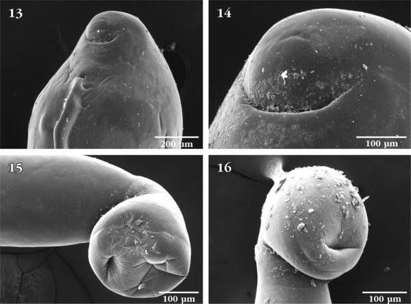
SEM of Neoechinorhynchus manubrianus: 13. Ventral view of the posterior end of a female specimen showing the position of the gonopore; 14. Enlargement of the posterior end of specimen in Fig. 13, showing the near terminal subterminal position of the crescent-shaped gonopore; 15. Ventro-lateral view of the bursa of a male specimen showing its bland appearance and articulation at an angle from the posterior trunk; 16. Near-ventral view of the same bursa in Fig. 15, showing its non-central orifice.
Current usage metrics show cumulative count of Article Views (full-text article views including HTML views, PDF and ePub downloads, according to the available data) and Abstracts Views on Vision4Press platform.
Data correspond to usage on the plateform after 2015. The current usage metrics is available 48-96 hours after online publication and is updated daily on week days.
Initial download of the metrics may take a while.