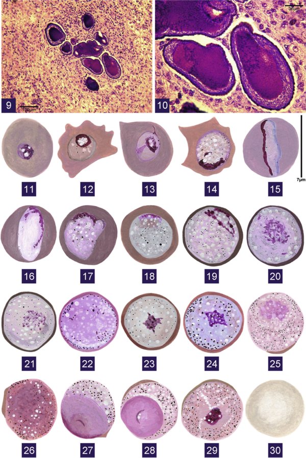
Figs 9–10 & 11–30.

Download original image
Fig. 9: microphotograh of schizonts of Hepatocystis levinei in liver sections from Pteropus alecto; Fig. 10: detail of Fig. 9; Figs 11-30: giemsa stain; Figs 11-24: drawings of gametocytes of Sprattiella alecto; Fig. 11: very young trophozoite; Figs 12-18: young trophozoites; Figs 18-21: immature microgametocytes; Fig. 22: mature microgametocyte; Figs 23-24: macrogametocytes; Figs 25-28: microgametocytes of Hepatocystis sp.; Fig. 29: Microgametocyte “en cocarde” of Hepatocystis levinei; Fig. 30: uninfected RBC
Current usage metrics show cumulative count of Article Views (full-text article views including HTML views, PDF and ePub downloads, according to the available data) and Abstracts Views on Vision4Press platform.
Data correspond to usage on the plateform after 2015. The current usage metrics is available 48-96 hours after online publication and is updated daily on week days.
Initial download of the metrics may take a while.


