| Issue |
Parasite
Volume 20, 2013
|
|
|---|---|---|
| Article Number | 27 | |
| Number of page(s) | 6 | |
| DOI | https://doi.org/10.1051/parasite/2013027 | |
| Published online | 29 August 2013 | |
Short Note
Schistosoma mansoni experimental infection in Mus spretus (SPRET/EiJ strain) mice
Infection expérimentale de Schistosoma mansoni chez la souris Mus spretus (souche SPRET/EiJ)
1
Laboratorio de Inmunología y Parasitología Molecular, CIETUS, Facultad de Farmacia, Universidad de Salamanca, 37008 Salamanca, Spain
2
Instituto de Investigaciones Biomédicas de Salamanca (IBSAL), Salamanca, Spain
3
Departamento de Estadística, Universidad de Salamanca, 37008 Salamanca, Spain
4
Centro de Investigación del Cáncer, Universidad de Salamanca-CSIC, 37008 Salamanca, Spain
* Corresponding Author: This email address is being protected from spambots. You need JavaScript enabled to view it.
Received:
9
June
2013
Accepted:
14
August
2013
Abstract
Most Schistosoma mansoni experimental infections are developed in several inbred strains of Mus musculus as definitive host. In contrast, Mus spretus is unexplored in Schistosoma infection studies. Mus spretus provides a high variation of immunological phenotypes being an invaluable tool for genetic studies and gene mapping. The aim of this study is to characterize hematological and immunological responses against Schistosoma mansoni infection in Mus spretus (SPRET/EiJ strain) vs. Mus musculus (CD1 strain) mice. Nine weeks after cercarial exposure, animals were perfused and the parasite burden was assessed. The parasitological data suggests that SPRET/EiJ mice tolerate higher parasite loads compared to CD1 strain. In addition, hematological parameters measured in Mus spretus group showed a significant increase in granulocytes population in early stages of infection compared to the CD1 cohort. Meanwhile, CD1 presented higher levels of lymphocytes and IgG1 in the late stages of S. mansoni experimental infection.
Résumé
La plupart des infections expérimentales à Schistosoma mansoni sont développées chez Mus musculus comme hôte définitif. Au contraire, Mus spretus est une espèce de souris inexplorée dans les infections expérimentales á Schistosoma. Mus spretus offre une grande variation de phénotypes immunologiques, ce qui est un outil essentiel pour les études génétiques et la cartographie des gènes. L’objectif de cette étude est la caractérisation de la réponse hématologique et immunologique contre l’infection à Schistosoma mansoni chez Mus spretus (souche SPRET/EiJ) comparée à Mus musculus (souche CD1). Neuf semaines après l’exposition aux cercaires, les animaux ont été perfusés et les paramètres parasitologiques ont été obtenus. Les données parasitologiques suggèrent que la souche SPRET/EiJ tolère des charges parasitaires plus élevées que la souche CD1. Les paramètres hématologiques mesurés chez SPRET/EiJ ont montré aussi une augmentation significative de la population des granulocytes dans les premiers stades de l’infection par comparaison à la cohorte CD1. Cependant, la souche CD1 a présenté des niveaux plus élevés de lymphocytes et IgG1 dans les stades tardifs de l’infection expérimentale à S. mansoni.
Key words: Schistosoma mansoni infection / Mus spretus / Immunological phenotypes / Hematological phenotypes
© L. Pérez del Villar et al., published by EDP Sciences, 2013
 This is an Open Access article distributed under the terms of the Creative Commons Attribution License (http://creativecommons.org/licenses/by/2.0), which permits unrestricted use, distribution, and reproduction in any medium, provided the original work is properly cited.
Introduction
Schistosomiasis remains one of the most important parasitic diseases affecting over 200 million human beings and causing 200,000 deaths per year [24]. However, the pathology caused by Schistosoma spp. infection varies widely depending on the intensity of infection and ecological factors. These issues contribute toward the differential global infection and mortality rates [2]. Furthermore, schistosomiasis susceptibility is influenced by multiple genes as well as by gene-gene and gene-environment interactions [6]. In experimental infections, inbreed mouse strains develop different degree of Schistosoma pathology; among these mouse strains, CBA/2J and C3H strains develop significantly higher hepatic pathology than C57BL/6J [5, 23]. At the late stages of experimental infections, the decrease of peripheral neutrophils is associated with an increase of lesion size and fibrosis in CBA mice, whereas those effects are minimal in C57BL/6 strain, indicating that neutrophils play a regulating role for granuloma formation [8]. Furthermore, enhanced neutrophil apoptosis has been reported in hepatosplenic schistosomiasis in humans [1]. Thus, identifying relevant hematological phenotypes involved in schistosomiasis susceptibility provides a useful insight into its pathogenesis in humans [3, 21].
Mus spretus have been the subject of various inquiries covering biometrical and morphological analyses [28]. Phylogenetic studies of mitochondrial D-loop sequences let to distinguish M. spretus from M. spicilegus and M. musculus as different species [11]. In fact, strains derived from wild M. spretus mice (i.e. SPRET/EiJ) show different divergent phenotypes and higher genetic variability than other common laboratory strains derived from M. musculus. Due to this phylogenetic difference, M. spretus mice have been useful for dissecting the genetic architecture of different complex traits including obesity, cancer, and infectious diseases [12, 22, 25, 26]. However, information about the susceptibility of M. spretus to experimental S. mansoni infection is not available. To determine whether there were specific variations in immunological and hematological responses against S. mansoni infection, we infected M. spretus mice (SPRET/EiJ), and compared their immunological response and infection parameters with those of Mus musculus (CD1) mice.
Materials and methods
Parasite and mice
Mus spretus (SPRET/EiJ) and Mus musculus (CD1) mice five-to six-week-old were purchased from the Jackson Laboratory and maintained in the Animal Facility at the University of Salamanca. All animals were treated according to the provisions of the current European law on animal experimentation. Cercariae of S. mansoni were obtained from infected Biomphalaria glabrata snails breeding in the Laboratory of Immunological and Molecular Parasitology, CIETUS, at the University of Salamanca. Forty mice (10♂ and 10♀ SPRET/EiJ and similarly 10♂ and 10♀ CD1) were included in the experiment. Each mouse was infected subcutaneously with 150 S. mansoni cercariae. Blood samples were collected at 0, 3, 6, and 9 weeks after infection. Animals were perfused at 9 weeks after infection and adult male and female parasites were counted with a dissecting microscope (10×) as previously described [20]. At the time of perfusion, small intestines and livers were collected and digested in 4% KOH for measuring the number of eggs deposited in these organs. Macroscopic lesions in the liver were quantified as granuloma affected surface per cm2 in each animal using the Image J software [18].
Hematological analysis and quantitation of serum immunoglobulins
Fifty microliters of mouse blood was collected in EDTA-coated tubes (Vacutainer®), then mixed and analyzed using the HEMAVET system®. The measurement of specific antibodies (IgG, IgG1, and IgG2a) against the Specific Worm Antigen Product (SWAP) was performed using an indirect ELISA. The Specific Worm Antigen Product (SWAP) was obtained as previously described [13]. The results were expressed as means of the optical density from all the animals of each group plus the standard error (SEM).
Data analysis
Statistical significance of parasitological data was analyzed by Kruskal-Wallis test. Differences with a p-value <0.05 were considered as statistically significant. All data were analyzed with the statistical software SPSS for Windows 11.5 (Lead Technologies), and visually displayed using the Lattice Package developed in R [17].
Results and discussion
Several studies have reported that Mus spretus are highly resistant mice to inflammation process [9, 14, 27]. Thus, Mus spretus have been proposed as useful model to identify genetic regions contributing to differences in immune response and inflammation. Therefore, the aim of the present study was to explore the hematological features and the immunophenotypes associated to resistance/susceptibility to schistosomiasis in SPRET/EiJ mice. The first finding related to the resistance/susceptibility to the experimental S. mansoni infection was the difference on infection rates. Our results indicated that SPRET/EiJ mice tolerate S. mansoni infection better than CD1 strain. Total worms recoveries were higher in SPRET/EiJ than in CD1 mice (Table 1); and trapped eggs in intestine and hepatic tissues were significantly higher in SPRET/EiJ too. We also found a higher percentage of macroscopic lesions caused by S. mansoni eggs in SPRET/EiJ mice compared with CD1 mice (Table 1). The evaluation of macroscopic granulomatous lesions was positively correlated with the intensity of infection useful for evaluating S. mansoni experimental infections [19]. Therefore, there was no apparent association between worm and tissue eggs burdens presented by SPRET/EiJ and its resistance to inflammation process. It should be noticed that the high intensity infection rate was also observed in C57BL/6 that is considered as resistant mouse strain to develop the severe form of schistosomiasis [16]. Concerning the effect of the mouse gender on parasitological data, we did not observe any statistical differences in any strain of mice.
Worm recovery, eggs counts in hepatic and intestinal tissues, and granulomatous lesions of Mus spretus (SPRET/EiJ strain) and Mus musculus (CD1 strain) mice.
In this study, we also analyzed the immunological and hematological profiles presented during S. mansoni infection in SPRET/EiJ and CD1 mice. The variables studied were white blood cell counts, and IgG1 and IgG2a isotype-specific subclasses. Regarding the humoral response, our results show a typical progressive enhancement on antibodies levels against the SWAP during S. mansoni experimental infection in both subtypes of immunoglobulin analyzed. Specifically, SPRET/EiJ mice showed an increase of IgG2a levels compared to CD1 at 9 weeks post-infection in both sexes (p < 0.05) (Figure 1A). On the other hand, female CD1 mice showed significant higher IgG1 levels at 3 and 6 weeks after infection compared with female SPRET/EiJ mice (p < 0.05). Furthermore, significant higher IgG1 levels were also found in CD1 male mice compared to their SPRET/EiJ counterparts at 6 weeks after infection (p < 0.05) (Figure 1B). These results suggested that CD1 mice would present a stronger Th2 immune response against S. mansoni compared to SPRET/EiJ mice.
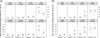 |
Figure 1. Boxplot graph displaying the levels of IgG2a (A) and IgG1 (B) along Schistosoma mansoni infection, according to sex and strain of mice. +p < 0.05. |
We hypothesized that the permissiveness of SPRET/EiJ mice to S. mansoni infection could be mediated by cell immune response in initial phase of infection. Therefore, we have also monitored blood cells parameters after 0, 3, 6, and 9 weeks during the S. mansoni infection in both mouse species. We noticed that SPRET/EiJ mice differ from CD1 in the hematological response. Interestingly, SPRET/EiJ presented high levels of peripheral blood granulocytes at 3 weeks after infection (Table 2). Blood granulocytes play a critical role in host defense mechanisms against invading pathogens; they are rapidly recruited to the infection site. In addition, these cell types are present in the initiation and maintenance of chronic allergic inflammation, and they could play a protective role in the immune response against S. mansoni [15]. In fact, published studies have shown that mast cells and eosinophils are able to produce IL-4 and modulate granuloma formation in S. mansoni infection [7, 10]. It has also been confirmed that SPRET/EiJ mice show a high level of resistance to infection of other pathogens like Salmonella enterica where macrophages and neutrophils also play a critical role [4]. Previous studies showed that SPRET/EiJ strain is also strongly resistant to inflammation induced either by cytokines or by bacterial products [27]. Regarding the hematological levels at 6 weeks after infection, we could observe that higher levels of neutrophils, eosinophils, and basophils continue to be associated to SPRET/EiJ strain, meanwhile, higher levels of lymphocytes populations were significantly associated to CD1 mice (p < 0.05) (Table 3). We also noted that total WBCs and lymphocytes populations were significantly decreased in SPRET/EiJ strain at 9 weeks after infection (p < 0.05). Therefore, despite attenuation of lymphocytes populations in SPRET/EiJ mice at the late stage of infection, SPRET/EiJ mice showed an enhanced tolerability to S. mansoni infection probably due to improved functions of the innate immune cells such as neutrophils in early time of S. mansoni infection. Finally, with respect the effect of mouse gender on hematological data, we did not observe any statistical significant differences in any species of mice.
Summary of granulocytes count in Mus spretus (SPRET/EiJ strain) and Mus musculus (CD1 strain) mice during Schistosoma mansoni infection.
Summary of blood parameters in Mus spretus (SPRET/EiJ strain) and Mus musculus (CD1 strain) mice during Schistosoma mansoni infection.
The present study highlights the importance of Mus spretus to unravel mechanisms of resistance/susceptibility against S. mansoni infection. The elevated neutrophil counts probably could be a reason for the tolerability of SPRET/EiJ mice against S. mansoni infection. Further analysis of the complex host response to S. mansoni infection, combined with the recent availability of the complete SPRET/EiJ genome sequence, will contribute to more understanding of the genetic control of host immunity against S. mansoni infection and the essential role of neutrophils in early immune defense mechanisms.
Acknowledgments
We gratefully acknowledge Dr. J. López-Abán for his technical assistance and Luis Miguel Rincón for reviewing the manuscript. This work was supported by the Program entitled “Plan propio de apoyo a la investigación 2013” of the University of Salamanca.
References
- Aref S, El Refaei MF, Sakrana M, El-Nemre H. 2004. Enhanced neutrophil apoptosis in neutropenic patients with hepatosplenic schistosomiasis: evidence of serum Fas ligand. Hematology, 9, 71–78. [CrossRef] [PubMed] [Google Scholar]
- Campino S, Kwiatkowski D, Dessein A. 2006. Mendelian and complex genetics of susceptibility and resistance to parasitic infections. Seminars in Immunology, 18, 411–422. [CrossRef] [PubMed] [Google Scholar]
- Cooke GS, Hill AV. 2001. Genetics of susceptibility to human infectious disease. Nature Reviews Genetics, 2, 967–977. [CrossRef] [PubMed] [Google Scholar]
- Dejager L, Pinheiro I, Bogaert P, Huys L, Libert C. 2010. Role for neutrophils in host immune responses and genetic factors that modulate resistance to Salmonella enterica serovar typhimurium in the inbred mouse strain SPRET/EiJ. Infection and Immunity, 78, 3848–3860. [CrossRef] [PubMed] [Google Scholar]
- Farah IO, Kariuki TM, King CL, Hau J. 2001. An overview of animal models in experimental schistosomiasis and refinements in the use of non-human primates. Lab Animal, 35, 205–212. [CrossRef] [Google Scholar]
- Flint J, Valdar W, Shifman S, Mott R. 2005. Strategies for mapping and cloning quantitative trait genes in rodents. Nature Reviews Genetics, 6, 271–286. [CrossRef] [PubMed] [Google Scholar]
- Gessner A, Mohrs K, Mohrs M. 2005. Mast cells, basophils, and eosinophils acquire constitutive IL-4 and IL-13 transcripts during lineage differentiation that are sufficient for rapid cytokine production. Journal of Immunology, 174, 1063–1072. [Google Scholar]
- Hirata M, Hara T, Kage M, Fukuma T, Sendo F. 2002. Neutropenia augments experimentally induced Schistosoma japonicum egg granuloma formation in CBA mice, but not in C57BL/6 mice. Parasite Immunology, 24, 479–488. [CrossRef] [PubMed] [Google Scholar]
- Hochepied T, Schoonjans L, Staelens J, Kreemers V, Danloy S, Puimège L, Collen D, Van Roy F, Libert C. 2004. Breaking the species barrier: derivation of germline-competent embryonic stem cells from Mus spretus x C57BL/6 hybrids. Stem Cells, 22, 441–447. [CrossRef] [PubMed] [Google Scholar]
- Mohrs K, Wakil AE, Killeen N, Locksley RM, Mohrs M. 2005. A two-step process for cytokine production revealed by IL-4 dual-reporter mice. Immunity, 23, 419–429. [CrossRef] [PubMed] [Google Scholar]
- Musser GG, Carleton MD. 2005. Superfamily Muroidea in Mammal species of the world: a taxonomic and geographic reference, Wilson Don E., Reeder Dee Ann M. Editors. The Johns Hopkins University Press: Baltimore. p. 894–1531. [Google Scholar]
- Nagase H, Mao JH, Balmain A. 1999. A subset of skin tumor modifier loci determines survival time of tumor-bearing mice. Proceedings of the National Academy of Sciences of the United States of America, 96, 15032–15037. [CrossRef] [PubMed] [Google Scholar]
- Pardo J, Carranza C, Turrientes MC, Pérez Arellano JL, López Vélez R, Ramajo V, Muro A. 2004. Utility of Schistosoma bovis adult worm antigens for diagnosis of human schistosomiasis by enzyme-linked immunosorbent assay and electroimmunotransfer blot techniques. Clinical and Diagnostic Laboratory Immunology, 11, 1165–1170. [PubMed] [Google Scholar]
- Quigley DA, To MD, Pérez-Losada J, Pelorosso FG, Mao JH, Nagase H, Ginzinger DG, Balmain A. 2009. Genetic architecture of mouse skin inflammation and tumour susceptibility. Nature, 458, 505–508. [CrossRef] [PubMed] [Google Scholar]
- Rumbley CA, Sugaya H, Zekavat SA, El Refaei M, Perrin PJ, Phillips SM. 1999. Activated eosinophils are the major source of Th2-associated cytokines in the schistosome granuloma. Journal of Immunology, 162, 1003–1009. [Google Scholar]
- Rutitzky LI, Mirkin GA, Stadecker MJ. 2003. Apoptosis by neglect of CD4+ Th cells in granulomas: a novel effector mechanism involved in the control of egg-induced immunopathology in murine schistosomiasis. Journal of Immunology, 171, 1859–1867. [Google Scholar]
- Sarkar D. 2008. Lattice: Multivariate Data Visualization with R. New York: Springer. [Google Scholar]
- Schneider CA, Rasband WS, Eliceiri KW. 2012. NIH Image to ImageJ: 25 years of image analysis. Nature Methods, 9, 671–675. [Google Scholar]
- Shariati F, Pérez-Arellano JL, Carranza C, López-Abán J, Vicente B, Arefi M, Muro A. 2011. Evaluation of the role of angiogenic factors in the pathogenesis of schistosomiasis. Experimental Parasitology, 128, 44–49. [CrossRef] [PubMed] [Google Scholar]
- Siles-Lucas M, Uribe N, López-Abán J, Vicente B, Orfao A, Nogal-Ruiz JJ, Feliciano AS, Muro A. 2007. The Schistosoma bovis Sb14-3-3zeta recombinant protein cross-protects against Schistosoma mansoni in BALB/c mice. Vaccine, 25, 7217–7223. [CrossRef] [PubMed] [Google Scholar]
- Smith PM, Shainheit MG, Bazzone LE, Rutitzky LI, Poltorak A, Stadecker MJ. 2009. Genetic control of severe egg-induced immunopathology and IL-17 production in murine schistosomiasis. Journal of Immunology, 183, 3317–3323. [CrossRef] [Google Scholar]
- Staelens J, Puimège L, Mahieu T, Pynaert G, Hochepied T, Vandenabeele A, Grooten J, Kontoyiannis D, Van Roy F, Kollias G, Libert C. 2004. Response of TNF-hyporesponsive SPRET/EiJ mice in models of inflammatory disorders. Mammalian Genome, 15, 537–543. [CrossRef] [Google Scholar]
- Stavitsky AB. 2004. Regulation of granulomatous inflammation in experimental models of schistosomiasis. Infection and Immunity, 72, 1–12. [CrossRef] [PubMed] [Google Scholar]
- Steinmann P, Keiser J, Bos R, Tanner M, Utzinger J. 2006. Schistosomiasis and water resources development: systematic review, meta-analysis, and estimates of people at risk. Lancet Infectious Diseases, 6, 411–425. [CrossRef] [Google Scholar]
- Stephan K, Smirnova I, Jacque B, Poltorak A. 2007. Genetic analysis of the innate immune responses in wild-derived inbred strains of mice. European Journal of Immunology, 37, 212–223. [CrossRef] [PubMed] [Google Scholar]
- To MD, Perez-Losada J, Mao JH, Hsu J, Jacks T, Balmain A. 2006. A functional switch from lung cancer resistance to susceptibility at the Pas1 locus in Kras2LA2 mice. Nature Genetics, 38, 926–930. [CrossRef] [PubMed] [Google Scholar]
- Turcotte K, Loredo-Osti JC, Fortin P, Schurr E, Morgan K, Gros P. 2006. Complex genetic control of susceptibility to Mycobacterium bovis (Bacille Calmette-Guerin) infection in wild-derived Mus spretus mice. Genes and Immunity, 7, 684–687. [CrossRef] [PubMed] [Google Scholar]
- Veyrunes F, Britton-Davidian J, Robinson TJ, Calvet E, Denys C, Chevret P. 2005. Molecular phylogeny of the African pygmy mice, subgenus Nannomys (Rodentia, Murinae, Mus): implications for chromosomal evolution. Molecular Phylogenetics and Evolution, 36, 358–369. [CrossRef] [PubMed] [Google Scholar]
Cite this article as: Pérez del Villar L, Vicente B, Galindo-Villardón P, Castellanos A, Pérez-Losada J & Muro A: Schistosoma mansoni experimental infection in Mus spretus ((SPRET/EiJ strain) mice. Parasite, 2013, 20, 27.
All Tables
Worm recovery, eggs counts in hepatic and intestinal tissues, and granulomatous lesions of Mus spretus (SPRET/EiJ strain) and Mus musculus (CD1 strain) mice.
Summary of granulocytes count in Mus spretus (SPRET/EiJ strain) and Mus musculus (CD1 strain) mice during Schistosoma mansoni infection.
Summary of blood parameters in Mus spretus (SPRET/EiJ strain) and Mus musculus (CD1 strain) mice during Schistosoma mansoni infection.
All Figures
|
Figure 1. Boxplot graph displaying the levels of IgG2a (A) and IgG1 (B) along Schistosoma mansoni infection, according to sex and strain of mice. +p < 0.05. |
| In the text | |
Current usage metrics show cumulative count of Article Views (full-text article views including HTML views, PDF and ePub downloads, according to the available data) and Abstracts Views on Vision4Press platform.
Data correspond to usage on the plateform after 2015. The current usage metrics is available 48-96 hours after online publication and is updated daily on week days.
Initial download of the metrics may take a while.